Tumor Rapid Fire Abstract
Tumor
Discussion
Sunday, May 5, 2024
1:59 PM - 2:09 PM CT
Location: S405a

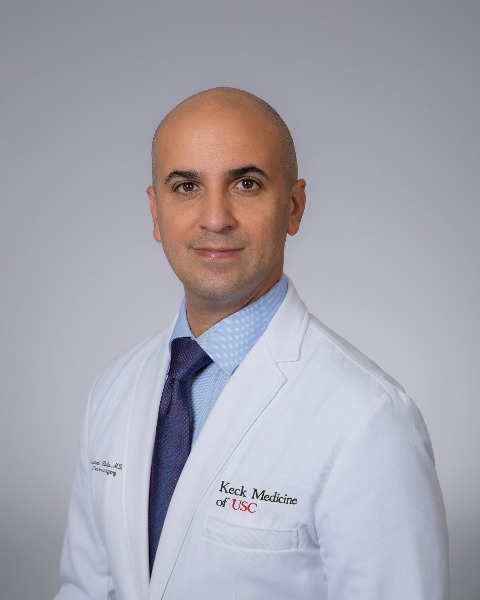
Gabriel Zada, MD
Neurosurgeon
University of Southern California
Los Angeles, CA, US
Moderator(s)
Disclosure(s):
Gabriel Zada, MD: Stryker Corp: Consultant (Ongoing)
Introduction: Language task-based fMRI can localize eloquent language areas preoperatively. In some left-dominant patients with left lesions, language dominance may translocate to the right hemisphere. However, the mechanisms behind this translocation are poorly understood. Graph theory describes functional network connectivity, illustrating complex network interactions between multiple language areas. We applied graph theory to task-based fMRI to examine language network reorganization in left-dominant patients with a left glioma.
Methods: We retrospectively identified 34 left-dominant patients with a left glioma and two fMRI scans (fMRI1 & fMRI2). In 8 patients, lateralization moved to the right hemisphere between the scans (reorganized group), while 26 stayed left-dominant (constant group). We constructed ROI-by-ROI correlation matrices to assess each group's connectivity at each scan. We computed graph theoretical measures, including clustering coefficients (CLC) and node centrality (NC), to examine connectivity differences between the two groups.
Results: At fMRI1, patients in the reorganized group displayed elevated functional connectivity in the right compared to the left hemisphere (CLC-left=0.97, CLC-right=0.88, p< 0.001). Between fMRI1 and fMRI2, each group's network restructured following a distinct pattern. Node centrality increased in the reorganized group (fMRI1: 0.18, fMRI2: 0.52, p< 0.001) but decreased in the constant group (fMRI1: 0.31, fMRI2: 0.19, p< 0.05). Network clustering decreased in the reorganized group (fMRI1: 0.50, fMRI2: 0.32, p< 0.001) but grew in the constant group (fMRI1: 0.35, fMRI2: 0.51, p< 0.001). Finally, the R-basal ganglia (∆NC=64, R-Broca's area (BA, ∆NC=54), and L-thalamus (∆NC=154) showed the highest restructuring in the reorganized group.
Conclusion : Left-dominant patients with left lesions who will become right-dominant already display elevated right hemisphere connectivity at baseline. The L-thalamus, R-BA, and R-basal ganglia may drive these lateralization changes. These findings lay the foundation for models predicting lateralization changes in glioma patients. Such models could guide neurosurgeons towards maximal tumor resection, lowering recurrence rates while preserving patients' language function.
